

Cornea and keratoconus assessment
Cornea and ocular-surface care addresses the clear front window of the eye, tear film and related structures. Examination distinguishes dryness, infection, inflammation, scarring and shape-related conditions such as keratoconus.
Symptoms and findings that need review
Persistent surface symptoms, pain, changing corneal shape or contact-lens difficulty deserve clinical assessment.
Persistent dryness or burning
Assessment can review tears, lids and environmental factors.
Pain, redness or light sensitivity
Marked or rapidly worsening symptoms need timely examination.
Changing corneal shape
Increasing astigmatism or suspected keratoconus may need mapping.
Contact-lens difficulty
Discomfort, reduced wearing time or recurrent redness should not be ignored.
How the consultation works
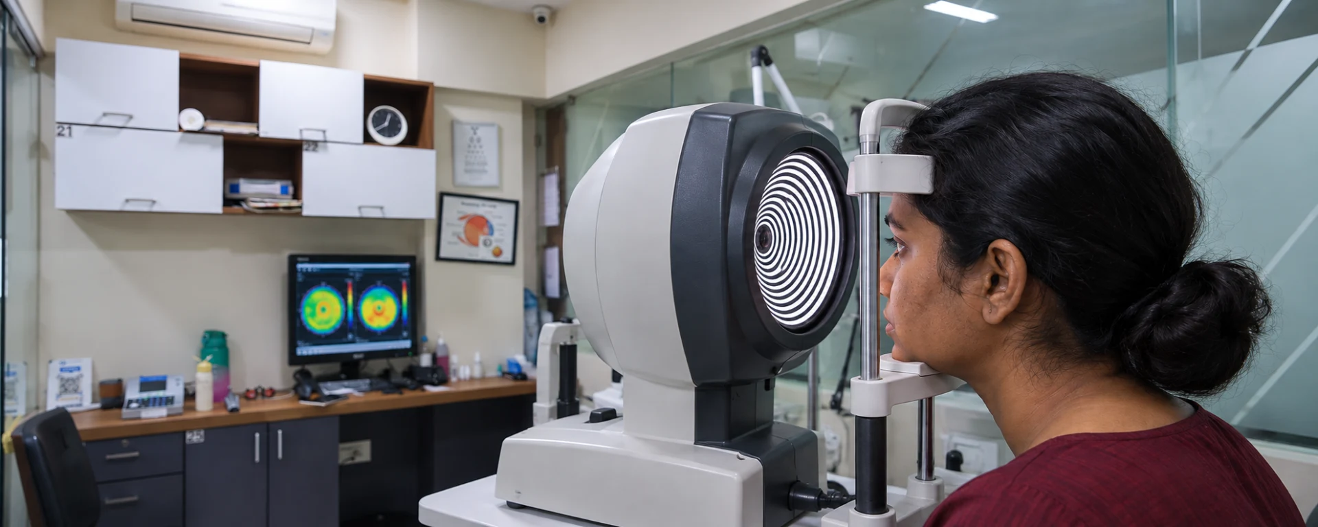
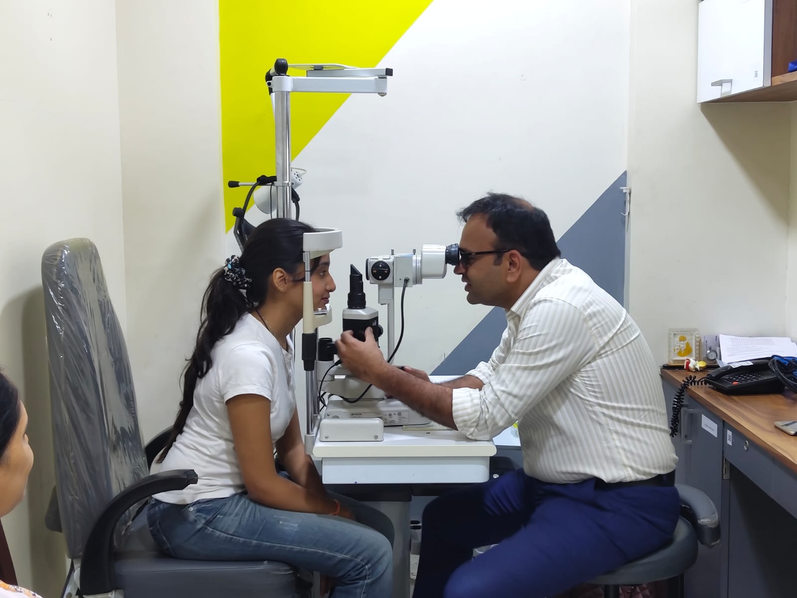
The ophthalmologist examines the cornea and tear film at the slit lamp, adding mapping, thickness measurements or staining when relevant.
- 01
Clarify symptoms, lens use, allergies and previous treatment.
- 02
Examine lids, tear film, conjunctiva and cornea.
- 03
Arrange topography or thickness measurements when needed.
- 04
Plan medical, optical, procedural or monitoring options.
Your Cornea & Keratoconus questions
Your ophthalmologist will explain what applies after examining your eyes.
No. Allergy, dryness, inflammation, injury and other causes can look similar.
It maps corneal shape and curvature to support assessment and lens fitting.
Serial measurements help determine stability or progression.
Sudden vision loss, severe eye pain, chemical exposure or significant eye injury needs urgent in-person medical attention.
.jpeg)
.jpeg)